
Receptor Occupancy
Receptor Occupancy and Target Engagement
A receptor occupancy assay measures how much of a target receptor a drug engages in tissue or in the living animal. It is the most direct readout of target engagement available in preclinical work, and it answers the question behind every dose decision:
“How much drug does it take to occupy a sufficient amount of receptors to matter?”
Gifford Bioscience runs ex vivo and in vivo receptor occupancy assays for central nervous system (CNS) and peripheral targets. We design the radiotracer assay, process and section the tissue, measure occupancy region by region where it matters, and report the dose-occupancy and concentration-occupancy relationships. It is these aspects that guide subsequent dose selection and PK/PD modelling.
Occupancy is measured by competition assays where the drug and a radiotracer compete for the same binding site. The more receptors the drug occupies, the less radiotracer that can bind. This fall in radiotracer signal, relative to vehicle-treated control tissue, is the receptor occupancy.
Occupancy and drug-induced effects are not always directly proportional (i.e., greater receptor occupancy elicits greater clinical effects), and assuming they are is a common way to misread dose-response outputs. Our previous work on the cannabinoid receptor, CB1, showed that the behavioural and neurochemical effects of cannabinoid agonists appear at very low occupancy, due to a large receptor reserve, which a functional assay alone would never consider. Receptor occupancy assays are a direct measure of what a drug dose engages.
Ex Vivo Receptor Occupancy
In an ex vivo receptor occupancy assay, the test compound is dosed in vivo, then tissue is collected at peak drug levels and frozen. We section the tissue on a cryostat and incubate the sections with a radiotracer for the target. Test compound that was dosed in vivo and has bound within the tissue will block a portion of radiotracer binding
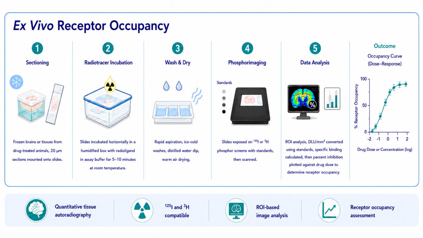
Quantitative autoradiography provides a direct measure of receptor distribution and drug-induced occupancy, linking target engagement to dose and supporting confident dose selection.
Because the radiotracer is applied to tissue sections, ex vivo occupancy resolves target engagement region by region. For CNS targets, that means reading occupancy separately across cortex, striatum, cerebellum and hippocampus, rather than averaging it across the whole tissue. This is where ex vivo occupancy earns its place: it shows not just whether a drug reaches the target, but also where.
In Vivo Receptor Occupancy
Where a suitable in vivo radiotracer exists, occupancy can be measured in the live rodent. The test compound is dosed, a radiotracer for the target is given intravenously and tissue is then collected at peak drug levels. We measure the reduction in radiotracer binding caused by the test compound, expressed as the ratio of binding in a receptor-rich region to a reference region.
Built on Validated Radiotracers
A receptor occupancy assay is only as good as the radiotracer behind it. Before an occupancy study, we at Gifford Bioscience characterise the radioligand using homogenate binding assays for affinity and binding-site density within the region of interest. These assays also confirm the radioligand specificity in the presence of selective blockers or, where available, the use of knockout tissue. Gifford Bioscience has developed and validated radiotracers for this purpose, including radioiodinated (through in-house iodination techniques) and tritiated ligands.
In Vivo Receptor Occupancy Case Study
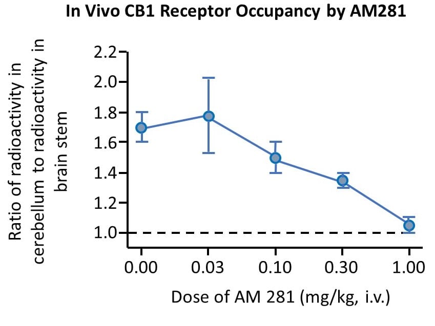
Occupancy of a cannabinoid drug AM281 at CB1 receptors, determined by inhibition of the corresponding radioiodinated tracer. The unlabelled drug reduced specific radiotracer binding by 50%, corresponding to approximately 50% receptor occupancy, at a dose of 0.3 mg/kg. Values are ratios of radioactivity between a CB1 receptor-rich brain region (cerebellum) and a lower-density region (brain stem). The dotted line indicates the level of non-specific binding.
Ex vivo Receptor Occupancy Case Study
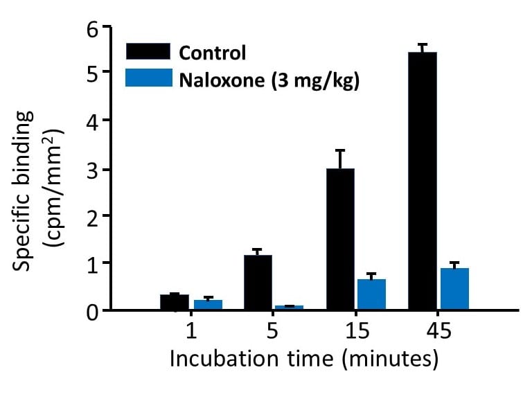
Using autoradiography, the radiolabelled opioid ligand [3H]DAMGO was incubated for different periods of time with striatal sections from an untreated rat (control) and from a rat dosed with the µ receptor antagonist naloxone (3 mg/kg), 20 minutes before sacrifice. Binding of [3H]DAMGO is reduced at all incubation time points in the sections from the treated animal, due to occupancy of µ receptors in the tissue by, naloxone.
Frequently Asked Questions about Receptor Occupancy
What is a receptor occupancy assay?
A receptor occupancy assay measures the proportion of a target receptor population that a drug binds at a given dose or exposure. It works by competition: the drug and a radiotracer compete for the same binding site, and the reduction in radiotracer binding reports the occupancy. It is the most direct measure of target engagement in preclinical drug development.
What is the difference between ex vivo and in vivo occupancy?
In an ex vivo assay the rodent is dosed with the test compound, tissue is collected and frozen, and the radiotracer is applied to tissue sections in the laboratory. Ex vivo works for any target within the sectioned tissue using a selective radiotracer and resolves occupancy by region. In an in vivo assay, the radiotracer and test compound are dosed and bind to the receptor in living tissue. In vivo needs a radiotracer with suitable kinetics in the rodent, but reports engagement under true physiological conditions.
Do you undertake the dosing?
No. Gifford Bioscience designs the radiotracer assay, processes and sections the tissue, measures occupancy and reports the dose-occupancy relationship. We coordinate tissue collection with the partner, so the assay and the dosing schedule line up.
Which receptors can you study?
Any receptor with a selective radiotracer can be studied. Gifford Bioscience has a depth of knowledge in CNS targets, including GPCRs, such as the, cannabinoid receptor, CB1, and opioid receptors, as well as transporters such as the dopamine transporter. Where a suitable radiotracer does not yet exist, we advise on its selection or development.
What radioisotopes are used?
Tritium [3H] and iodine-125 [125I] are used for ex vivo work on tissue sections. In vivo occupancy uses a radiotracer chosen for the target, which may be labelled with iodine-125.
Why measure occupancy if I already have a functional readout?
Because occupancy does not always equal drug effect. For some targets most of the receptor population must be engaged before an effect appears; for others, such as the CB1 cannabinoid receptor, an effect is produced at very low occupancy because of a large receptor reserve. Measuring occupancy directly tells you how much target a dose engages, which is what dose selection and PK/PD modelling depend on.
Can occupancy be resolved by brain region?
Yes, in the ex vivo format. Because the radiotracer is applied to tissue sections, occupancy is quantified separately in regions such as cortex, striatum, cerebellum and hippocampus. This shows whether a drug engages its target uniformly or preferentially in particular regions, which a homogenate measurement cannot.
How does receptor occupancy relate to PET?
PET measures occupancy non-invasively in living animals and humans, but with limited spatial resolution. Ex vivo and in vivo occupancy assays on tissue use higher-resolution methods and a matched radiotracer and are often run in the preclinical phase to support and interpret later PET studies of the same target.
What do you need from me to start?
The target receptor, the test compound, and the intended dosing route and schedule. From there we advise on the assay format, the radiotracer and the controls, coordinate tissue collection with the dosing partner, run the assay and report occupancy against dose or exposure.
Get in Touch



Copyright © 2026 Gifford Bioscience Limited
Privacy Policy