
Receptor Autoradiography
Receptor Autoradiography Services for Receptor Pharmacology
Gifford Bioscience specialize in receptor autoradiography to visualise and quantify the distribution of target receptors in human or animal tissue.
Receptor autoradiography is a quantitative imaging technique used to identify and map binding sites for a labelled drug in a tissue or organ section. Autoradiography preserves the spatial architecture that homogenate binding assays do not, providing region-by-region resolution of receptor location and density.
At Gifford Bioscience, receptor autoradiography is run on cryostat-cut sections of frozen human or untreated animal tissue. We offer in vitro autoradiography for receptor characterisation and competition studies, and ex vivo autoradiography for receptor occupancy determination from drug-treated animals.

We use Bright cryostat for precision sectioning of fresh-frozen tissue, enabling the thin, well-preserved sections essential for high-resolution receptor autoradiography.
What Is Receptor Autoradiography Used For?
In vitro autoradiography. Using sections cut from frozen untreated human or animal tissue, in vitro autoradiography is used to visualise and quantify the distribution of the target receptors in the tissue, through the binding of a radiolabeled drug. In vitro autoradiography can also identify off-target binding sites, by revealing residual binding in knockout animal tissue, or in regions where the target receptor is not expressed.
Ex vivo autoradiography, performed on tissue from drug-treated animals, determines the receptor occupancy of the drug at the time of tissue collection. Because the spatial distribution is preserved, quantification can be limited to only regions of interest / regions of high receptor expression.
For human tissue use, ethics committee approval is required. Gifford Bioscience manages the application process to obtain the tissue from an accredited UK tissue bank.
Example Data
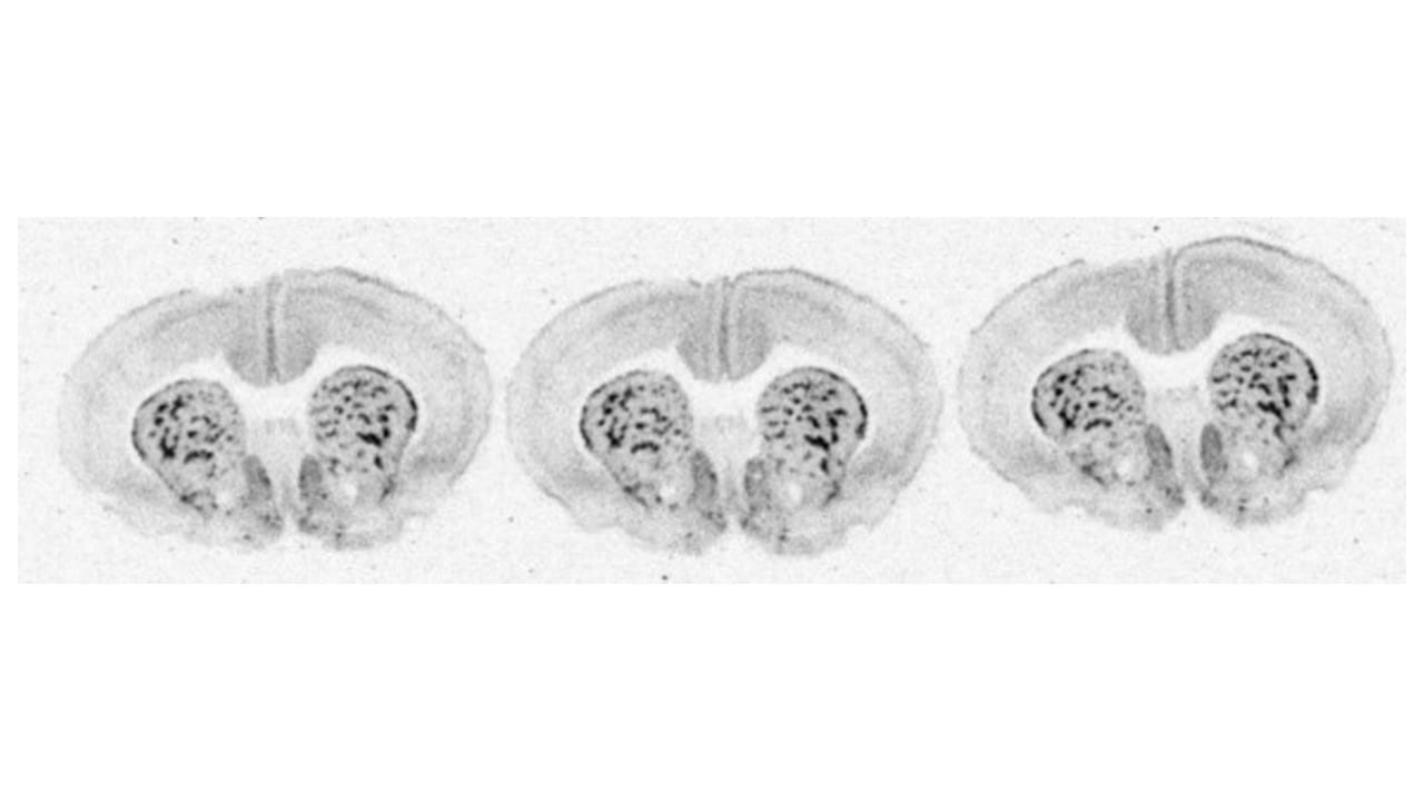
Binding of [3H]DAMGO to µ opioid in coronal sections at the level of the corpus striatum in a rat brain.
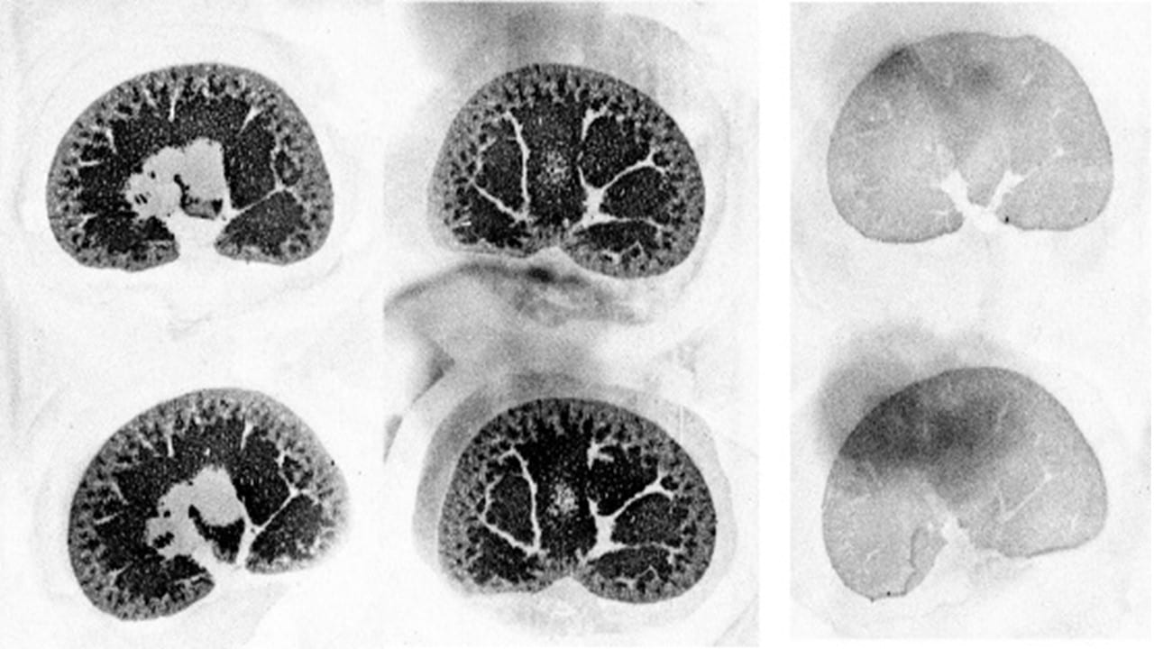
Binding of [3H]LRRK2-IN-1 to the LRRK2 enzyme in rat kidney. Sections in the centre and to the left are total binding. The two sections on the right are non-specific binding, defined using unlabeled LRRK2-IN-1 (10 µM).
When to Choose Autoradiography over Homogenate Radioligand Binding?
Homogenate radioligand binding is the right tool for affinity, density and competition measurements, averaged across a tissue. Receptor autoradiography is the right tool when spatial information matters, such as:
- regional receptor distribution mapping;
- identification of off-target binding sites;
- ex vivo receptor occupancy;
- work in low-abundance regions where homogenate dilution would lose signal;
- translational studies, where preserving anatomy is essential for read-across to in vivo imaging.
- The two approaches can be used in combination, with homogenate binding establishing affinity and autoradiography confirming spatial selectivity.
Autoradiography Protocol
Cryostat-cut tissue sections are incubated with the radiolabelled drug until equilibrium is reached. The sections are washed in fresh buffer to remove unbound radiotracer, dried and imaged using quantitative phosphorimaging. Regions-of-interest (ROIs) are then drawn over each imaged section and radioactivity levels are measured. Even smaller regions, down to 0.1 to 0.2 mm, can be quantified.
Non-specific binding is defined on adjacent sections using an excess of unlabelled competitor. Specific binding is calculated by subtraction. Calibrated standards can be used to convert phosphorimage signal to fmol of bound radioligand per mg of tissue.
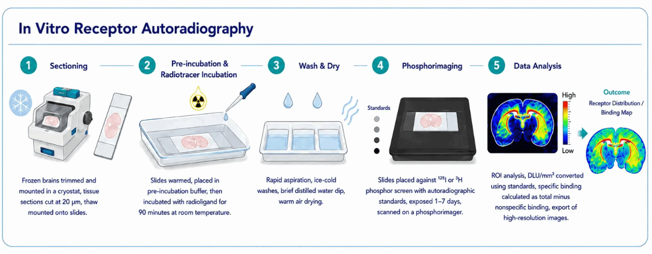
The Gifford autoradiography workflow, from cryostat sectioning through radioligand incubation, washing, phosphorimaging and ROI-based quantification.
Applications across Target Classes
Receptor autoradiography is applicable in situations where the density of target receptor is sufficiently high to visualize the receptors in the cryostat-cut sections. Applications include many common GPCRs, ligand-gated ion channels, enzymes and transporters, as well as certain pathologically aggregated proteins such as tau, amyloid-β and α-synuclein. The same workflow applies in each case, with the radioligand and incubation conditions tailored to the target.
Frequently Asked Questions about Receptor Autoradiography
What radioisotopes are used?
Gifford Bioscience has significant experience using Tritium [3H], Iodine-125 [125I], Sulphur-35 [35S] and other common laboratory radioisotopes.
Can autoradiography detect off-target binding?
Yes. Residual binding in knockout animal tissue, or in regions where the target receptor is not expressed, indicates binding to a non-target site.
Can receptor autoradiography be used for ion channels?
Yes. Both ligand-gated and voltage-gated ion channels can be mapped by autoradiography using subtype-selective radioligands, provided they have sufficient abundance in the tissue.
Examples include:
- nicotinic acetylcholine receptors using tritiated epibatidine or α-bungarotoxin;
- NMDA receptors using [3H]MK-801;
- GABA-A receptors using [3H]muscimol or [3H]flunitrazepam;
- voltage-gated sodium channels using [3H]saxitoxin.
The same protocol applies, with incubation conditions, buffer composition and non-specific binding controls optimised for the channel class.
Can autoradiography be used for tau, amyloid-β and other protein aggregates?
Yes. Autoradiography on post-mortem human brain sections is a common step in the validation of PET tracers for tau, amyloid-β and α-synuclein. The advantage of working with post-mortem tissue over recombinant proteins is that the aggregates reflect those in an actual disease state, rather than an in vitro produced synthetic aggregate.
What tissues can be used?
Frozen brain, kidney, lung, liver, heart and peripheral tissue from rodents, NHP and human donors. Human tissue is obtained from an accredited UK tissue bank following ethics committee approval, which Gifford Bioscience coordinates.
How is autoradiographic data quantified?
Phosphor screen signal is converted to fmol of bound radioligand per mg of tissue using co-exposed calibrated standards. Regions-of-interest are drawn over anatomically defined areas and reported as mean signal with non-specific binding subtracted. Data can be expressed as receptor density, percentage of control binding or percentage occupancy as needed.
Can binding kinetics be studied by autoradiography?
Yes, but homogenate aggregates would be more conventional for this.
How does autoradiography compare with PET and SPECT?
PET and SPECT are non-invasive in vivo techniques used in living animals and humans, but with limited spatial resolution. Autoradiography is performed on tissue sections ex vivo, with much higher spatial resolution, and is often used to validate or interpret in vivo imaging data with a matched radioligand.
Get in Touch



Copyright © 2026 Gifford Bioscience Limited
Privacy Policy